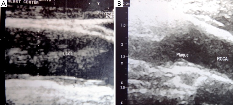
Figure 2.

(A) Longitudinal view of LCCA with 1.5 mm plaque at carotid body in 39 years old hypertensive and smoker male patient. Coronary angiography revealed total occlusion of mid LAD; (B) longitudinal view of RCCA with 1.3 mm plaque at carotid bulb in 33 years old normotensive, non-smoker female patient, presented with the history of acute myocardial infarction and coronary angiography revealed total occlusion of proximal LAD, who underwent primary PCI. LCCA, left common carotid artery; LAD, left anterior descending; RCCA, right common carotid artery; PCI, percutaneous coronary intervention.
