
Figure 4.
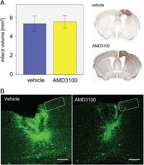
Effect of AMD3100 treatment on infarct size and accumulation of green fluorescent protein (GFP)-positive cells in the ischemic territory. (A) Infarct volumes in mice treated with saline (n = 9) or AMD3100 (n = 9) displayed as means ± SEM on day 14 after photothrombosis. Representative coronal sections of a vehicle- and AMD3100-treated animal are shown to the right. (B) Accumulation of GFP+ cells in the ischemic territory in a vehicle- and an AMD3100-treated CX3CR1+/GFP mouse. Scale bars: 200 μm. The frame illustrates the area analyzed for GFP+ cells by 2-photon-laser-microscopy.