
Figure 6.
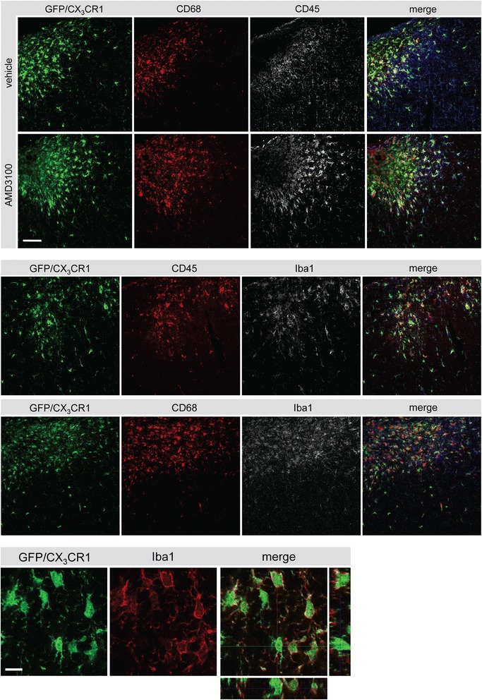
Phenotype of green fluorescent protein (GFP)-positive cells in the ischemic territory. Upper panels: colocalization of CD68 (Cy3, red), GFP (green, CX3CR1) and CD45 (white, Cy5) in the ischemic territory of a vehicle- and AMD3100-treated mouse after photothrombosis (PT). Middle panels illustrates the expression of ionized calcium binding adaptor molecule 1 (Iba1) in CD68/GFP- and CD45/GFP-positive cells. Scale bar: 50 μm. Lower panel: colocalization of GFP (green) with Iba1 (Cy3, red) in the ischemic core after PT. Scale bar: 10 μm.