
Figure 3.
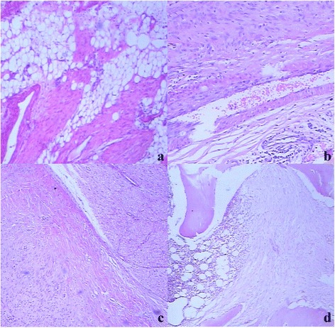
Histological features of postoperative recurrent desmoid tumors in the right forearm of a 15-year-old man (arrows) (a–d). (a) Lesions with adipose tissue involvement (HE × 40). (b) Desmoid tumors around vessels could not invade into the vessel wall to form tumor thrombus (HE × 40). (c) Desmoid tumors invaded into the connective tissue and perineurium around nerve tissue (HE × 40). (d) Desmoid tumors with bone involvement penetrated into the periosteum and cortical bone and invaded into the bone marrow cavity along the bone trabecula (HE × 40).