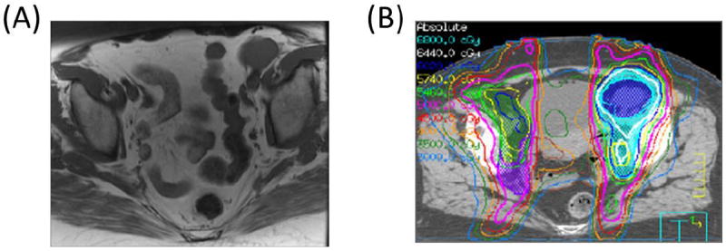
Fig. 2.

The location of the involved PLNs is important when definitive treatment options are being considered. (A) Axial T1 magnetic resonance imaging scan shows a large left distal external iliac lymph node measuring approximately 3.5 cm in maximal dimension. (B) This node is sufficiently far from critical structures that a simultaneous integrated boost and sequential boost to 64 Gy could be safely delivered. This patient is currently without evidence of disease 7.7 years after RT, with normal bladder and bowel function.
