
Fig 1.
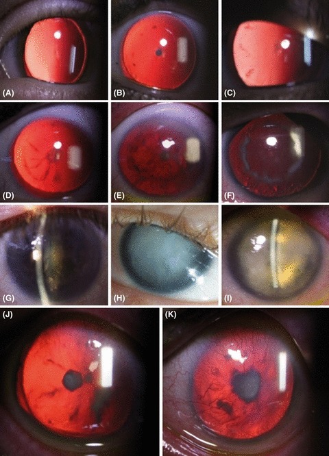
Developmental patterns of cataract in aniridia patients. (A) Early stage discrete posterior subcapsular cataract. (B–E) The posterior subcapsular cataract is denser. A posterior subcapsular opacification develops on the capsule in the mid-periphery as tiny flecks (B), or short, radially oriented opacities (C). (D–F) Radial opacities extend towards to posterior pole, and a mid-peripheral ring of opacification of varying density is present. Opacification always remained subcapsular. Late lens changes included yellow and turbid nuclear cataract (G), mature cataract (H) and hypermature cataract (I). (J) Anterior polar cataract in combination with a posterior polar cataract in one patient. (K) One patient presented with anterior polar cataract only.