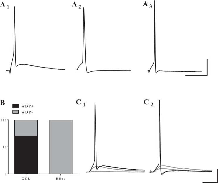
Fig. 5.
Cells in the GCL exhibit two distinct single AP waveforms in response to short depolarizing pulses, while hilar DGCs all exhibit similar waveforms. A: example traces of APs elicited by direct, somatic current injection. Cells in the GCL display AP traces: with an afterdepolarization (ADP; A1), or without an ADP (A2). A3: hilar cells only exhibit APs without an ADP. B: distribution of cells that fired each type of AP classified by location. The majority of cells in the GCL fired APs with an ADP (25/36), while none of the 7 hilar cells exhibited an ADP. C: a subset of cells in both the GCL and hilus could be driven synaptically to fire single APs. Of these cells, some fired an AP with ADP (C1), and others fired an AP without ADP (C2). Scale bars: 25 ms and 20 mV.

