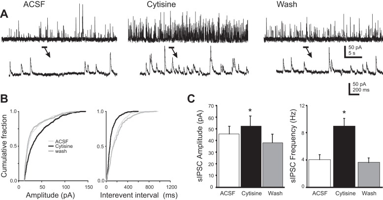
Fig. 4.
Effect of cytisine on spontaneous (s)IPSCs in a recorded dorsal motor nucleus of the vagus (DMV) neuron. A: recording of sIPSCs and response to bath-applied cytisine (100 μM) in a DMV neuron. Left: trace showing continuous recording in control ACSF. Center: the same cell in the presence of cytisine. Right: the same recording after 15-min washout to control ACSF. Arrows point to temporally expanded portions of each trace. B: cumulative probability plots for the traces shown in A. Cytisine increased both amplitude and frequency of sIPSCs in this recording (P < 0.05; Kolmogorov-Smirnov test). C: plots of mean amplitude (left) and frequency (right) for sIPSCs recorded for 7 DMV neurons, respectively. *Significant differences from control ACSF (P < 0.05).

