Fig. 3.
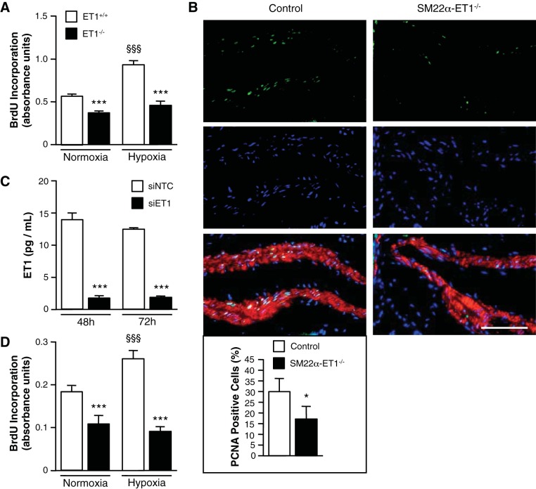
Loss of SMC ET-1 inhibits the hypoxia-induced proliferative response. A: loss of ET-1 significantly decreases mPASMC proliferation under both normoxic (21% O2) and hypoxic (5% O2) conditions. Cell proliferation was measured by bromodeoxyuridine (BrdU) incorporation assay after 24 h. ***P < 0.001, ET-1−/− vs. ET-1+/+; §§§P < 0.001, hypoxia vs. normoxia. B: representative images of proliferating cell nuclear antigen (PCNA)-expressing cells show a decrease in SMC proliferation in PA of SM22α-ET-1−/− mice exposed to hypoxia (3 wk, 10% O2) (right). PCNA, green; Hoechst (nuclei), blue; α-smooth muscle actin, red. Magnification ×400, scale bar = 50 μm. Percentage of PASMC ≥100 μm in diameter positive for PCNA expression. Graph represents the means ± SE of the number of PCNA-positive SMC/the total number of SMC per PA; n = 3 for each genotype with a minimum of 500 cells counted per mouse. *P < 0.05. C: ET-1 knockdown with siRNA in human PASMC (hPASMC) as assessed by ELISA. ***P < 0.001, siRNA specific for human ET-1 (siET-1) vs. scrambled nontargeted control siRNA (siNTC). D: loss of ET-1 significantly decreases hPASMC proliferation under both normoxic and hypoxic conditions. hPASMC were transfected with siNTC or siET-1. Cell proliferation was measured after 24 h by BrdU incorporation assay. ***P < 0.001, siET-1 vs. siNTC; §§§P < 0.001, hypoxia vs. normoxia.

