Fig. 5.
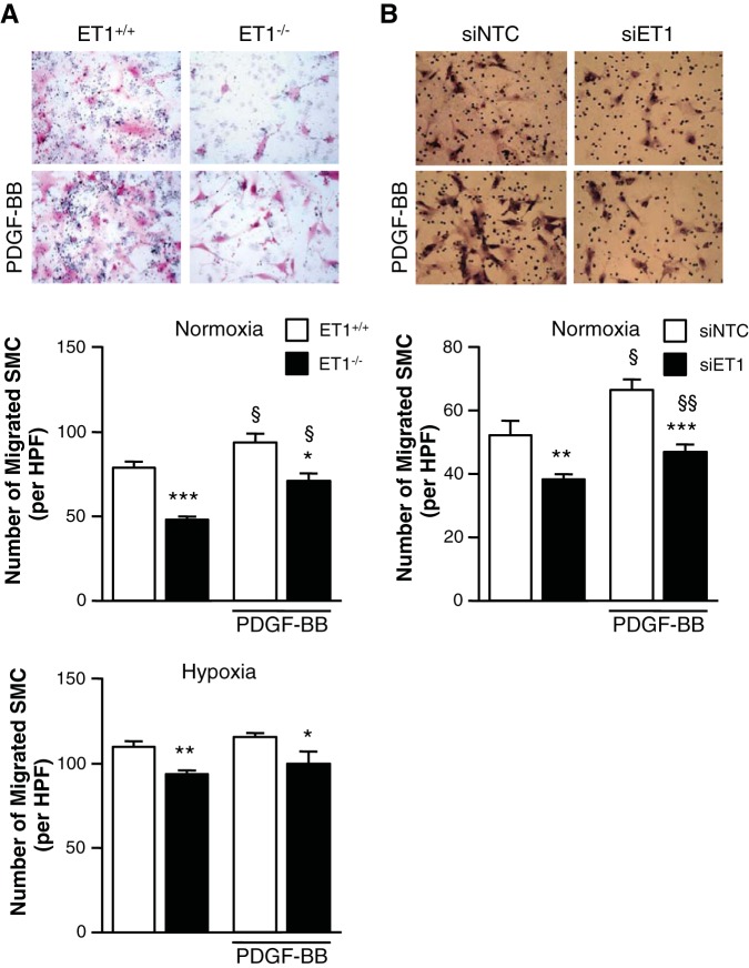
Loss of ET-1 inhibits SMC migration. A: mPASMC (ET-1+/+ and ET-1−/−) migration was assessed by modified Boyden Chamber assay. SMC were stimulated with or without 10 ng/ml PDGF-BB for 24 h. Representative images show that the loss of ET-1 (ET1−/−) inhibits SMC migration (right) independent of PDGF-BB stimulation (bottom). Graphs represent the means ± SE of the number of SMC per high-powered field (HPF) from 10 random fields per sample. *P < 0.05, **P < 0.01, ***P < 0.001, ET-1−/− vs. ET-1+/+; §P < 0.05, PDGF-BB vs. untreated. B: hPASMC migration was assessed by modified Boyden Chamber assay. SMC were transfected with siNTC or siET-1 and then stimulated with or without 10 ng/ml PDGF-BB for 24 h. Representative images show that the loss of ET-1 (siET-1) inhibits SMC migration (right) independent of PDGF-BB stimulation (bottom). Graph represents the means ± SE of the number of migrated SMC per HPF from 10 random fields per sample. Magnification ×200. **P < 0.01, ***P < 0.001, siET-1 vs. siNTC; §P < 0.05, §§P < 0.01, PDGF-BB vs. untreated.

