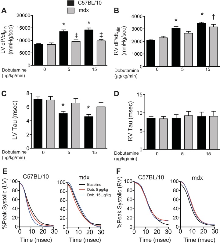
Fig. 7.
Evidence of disrupted relaxation in the mdx heart. The C57BL/10 left ventricle shows robust increases in relaxation in response to dobutamine documented as increased rate of relaxation (A) and isovolumic relaxation time constant, tau (C and E). In both of these measures of relaxation the mdx heart is not affected by dobutamine. The right ventricle demonstrates a dobutamine-induced increase in the rate of relaxation (B), but there is no dobutamine-induced decrease in tau (D and F). *P < 0.05, difference from baseline C57BL/10 data. ‡P < 0.05, difference from C57BL/10 at that dose. †P < 0.05, difference from baseline mdx.

