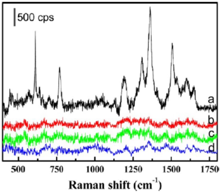
Figure 4.
SERS spectra of the MCF-7 cells with and without binding of the Rh6G-labeled aptamer-Ag-Au nanostructures (spectra a and b, respectively). SERS spectra of the HepG2 and MCF-10A (spectra c and d, respectively), after being incubated with the Rh6G-labeled aptamer-Ag-Au nanostructures, also are shown. Reprinted with permission form ref. 51. Copyright (2012) American Chemical Society.

