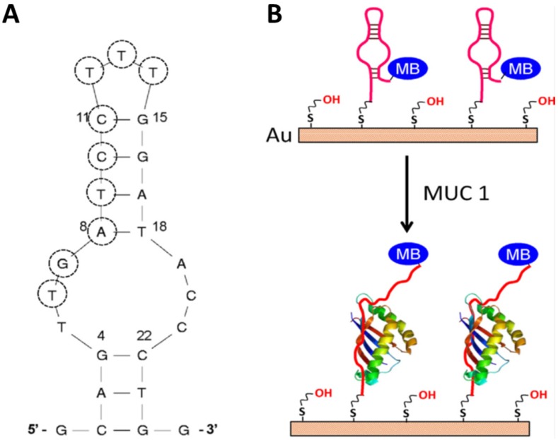
Figure 6.
The secondary structure of the S3.1/S2.2 anti-MUC1 DNA aptamer (A) and (B) the possible conformational change of MB-anti-MUC1-aptamer (immobi-lized on gold electrode) upon binding MUC1. Reprinted with permission from ref. 56. Copyright (2013) Elsevier.

