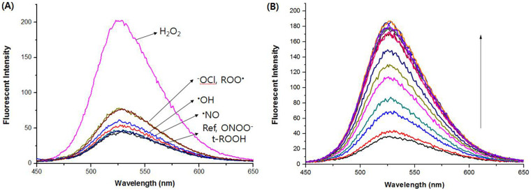
Figure 3.
(A) Fluorescence spectra of probe 1 (2 μM) with various ROS (200 μM) and (B) Fluorescent titration of probe 1 (2 μM) upon addition of H2O2 (0–100 eq.) in PBS (pH 7.4) solution containing 1% DMF after incubation for 2 h at 25°C at an excitation wavelength of 405 nm, and excitation and emission slit widths of 3 and 5 mm, respectively.

