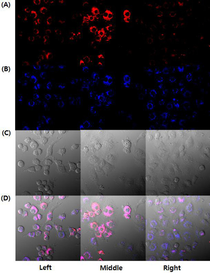
Figure 5. Probe 1 was localized to lysosomes in RAW 264.7 cell.
Confocal microscope images of probe 1 on the endogenous H2O2: (A) Probe 1 (red, ex405/em490–590 nm); (B) LysoTracker Blue DND-22 (blue, ex405/em430–455 nm); (C) Bright field (gray); (D) Overlay of (A) and (B) (purple). Left: No treatment; Middle: 1 μg/mL PMA (Phorbol 12-myristate 13-acetate), 1 h; Right: 1 μg/mL PMA, 1 h and 100 μM TEMPO (2,2,6,6-tetramethylpiperidine-1-oxyl, ROS scavenger), 1 h. (For producing endogenous H2O2, the RAW 264.7 cells were treated with PMA. All cells were stained with 5 μM probe 1, 100 nM Lysotracker for 30 min. Scale bar: 10 μm.).

