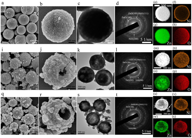
Figure 4.
The SEM (a–b, i–j and q–r), TEM (c, k and s) micrographs and SAED patterns (d, l and t) of ZnO-NiO solid, yolk-shell and hollow hybrid microspheres, respectively. HAADF STEM images (e, m and u) and the elemental mappings of Ni (f, n and v), Zn (g, o and w) and O (h, p and x) of ZnO-NiO solid, yolk-shell and hollow hybrid microspheres, respectively.

