
Figure 1.
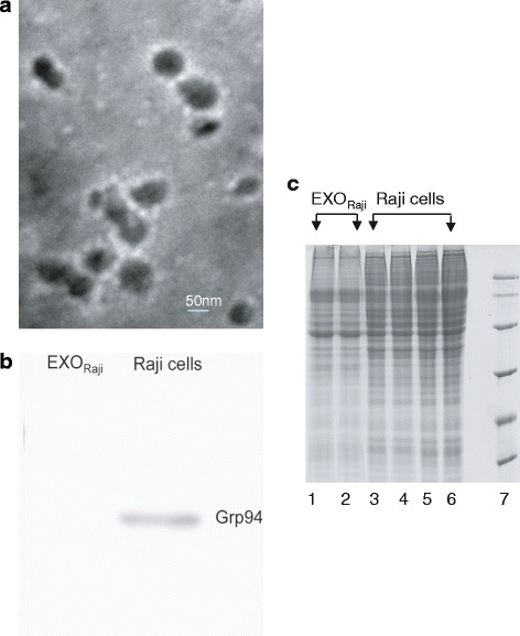
Morphology of EXO Raji and one-dimensional electrophoresis of samples from EXO Raji and Raji cells. a. Transmission electron microscope image of EXORaji (×100 K). b. Western blot analysis demonstrating the absence of Grp94 in EXORaji. c. Protein samples from EXORaji and Raji cells were separated by SDS-PAGE for further MS analysis. Lanes 1 and 2 represent Raji cell-derived exosomes, and lanes 3 to 6 represent Raji cells.