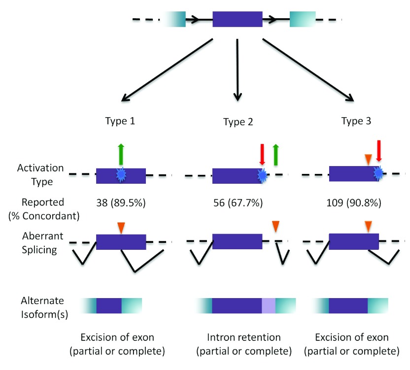
Figure 4. Outcomes of cryptic splicing mutations.
A prototypical internal exon (in purple) with flanking exons (in blue); introns are represented by black solid, and dashed lines (top). The three types of cryptic splice site activation are then illustrated. Type 1 cryptic splice site activation (left) is caused by the activation (green arrow) of a cryptic site by strengthening a pre-existing site, or by creating a novel splice site (blue). Type 2 (middle) results from the simultaneous weakening or abolition (red arrow) of the natural splice site while strengthening or creating (green arrow) a cryptic site. Type 3 (right) involves the activation of a pre-existing cryptic site due to the weakening or abolition of the natural splice site (indicated by orange triangle). The number of cases that have been reported in the literature that have been analyzed by IT for each type is indicated, with the percent accuracy in parentheses. The bottom row represents the resulting mRNA structure due to the activated cryptic splice site.

