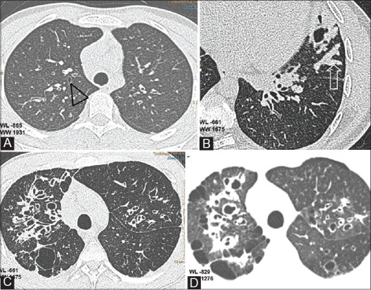
Figure 2(A-D).

Bronchiectasis and related findings. (A) Axial CT shows early bronchiectatic changes in right upper lobe bronchus (black triangle) (B)Mucus plug in lingular subsegment (open arrow) (C) Extensive bronchiectasis in right upper lobe with saccular cavities posteriorly (D) Thin section with soft algorithm (WL -829, WW 1276),reveals pulmonary aeration and bullous changes in the upper lobes with greater detail
