
Figure 1(A and B).
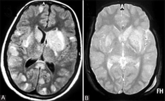
(A) Axial FLAIR and (B) gradient echo (GRE) MR images at the level of basal ganglia show multifocal FLAIR hyperintense lesions at gray-white matter junction and in bilateral basal ganglia. None of the lesions shows evidence of hemorrhage (B)