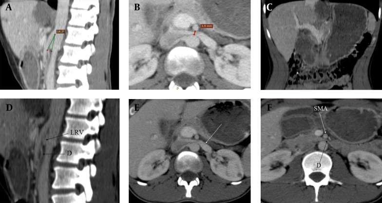
Figure 1. Superior Mesenteric Artery Syndrome Accompanying With Nutcracker Syndrome: CT Findings.
A) Sagittal CT scan showing the reduction of the angle (14 30) between the abdominal aorta and superior mesenteric artery. B) Axial CT scan showing decreased aortomesenteric distance (3, 9 mm). C) Coronal CT scan showing dilatation of the stomach and proximal duodenum. D) Sagittal CT scan of abdomen showing duodenal compression between the abdominal aorta and superior mesenteric artery (D: Duodenum, LRV: Left renal vein). E) Axial CT scan of abdomen demonstrating duodenal compression between the abdominal aorta and superior mesenteric artery (D, Duodenum; SMA, Superior mesenteric artery). F) Axial CT scan showing dilated left renal vein owing to superior mesenteric artery compressing (white arrow).

