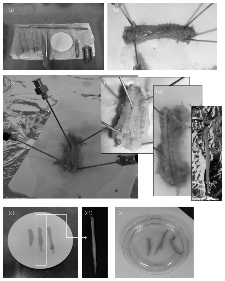
Figure 1.
Illustration of sequential steps of organotypic spinal cord slices preparation. All needed for preparation tools (a) and the main steps of the spinal cord isolation were photographed. The incision of the entire spinal cord block from the dorsal side (b). Fixation with syringe needles spinal column (c). Progressive steps of spinal cord isolation (c1–3). The dissection of white spinal cords (d) and its magnification (d1). The transfer of spinal cord slices into the Millicell-CM (Millipore) membranes (e).

