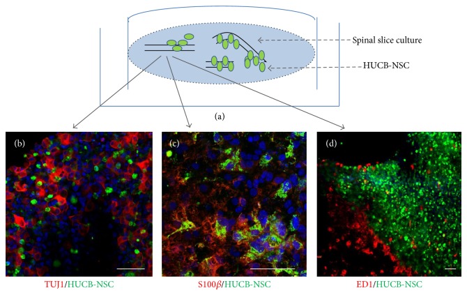
Figure 2.
Direct transplantation of HUCB-NSC into the spinal cord organotypic slice culture, an example of immunohistochemistry experiment. The cells were traced with CMFDA for their identification after engraftment. The transplanted cells (green) migrated inside the slice and spread out between the host neurons ((b); TUJ1, red). Three weeks after transplantation part of the transplanted cells expressed the astrocyte marker S100β ((c); red). The local immune response to xenografts was moderate ((d); ED1, red). Cell nuclei (blue) were stained with Hoechst 33258. Scale bar is 50 μm.

