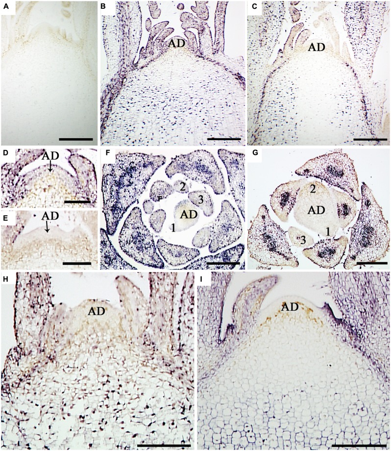
FIGURE 2.
In situ localization of CSVd in SAM of the CSVd infected Argyranthemum plants. (A) Longitudinal sections of healthy shoot tips of ‘Border Dark Red’. (B) Longitudinal section of CSVd-infected shoot tip of ‘Yellow Empire’. (C) Longitudinal section of CSVd-infected shoot tip of ‘Border Dark Red’. (D) Longitudinal section of CSVd-infected SAM of ‘Yellow Empire’ [higher magnification of the SAM in (B)]. (E) Longitudinal section of CSVd-infected shoot apical meristem (SAM) of ‘Border Dark Red’ [higher magnification of the SAM in (C)]. (F) Cross section of CSVd-infected shoot tip of ‘Yellow Empire’. (G) Cross section of CSVd-infected shoot tip of ‘Border Dark Red’. (H) Longitudinal section of CSVd-infected shoot tip of ‘Butterfly’. (I) Longitudinal section of CSVd-infected shoot tip of ‘Border Pink’. AD indicates apical dome (AD), and 1, 2, 3 indicates first, second, and the third leaf primordia, respectively. Scale bars in (A, B, C, H, I) are 310 μm; scale bars in (D–G) are 100 μm.

