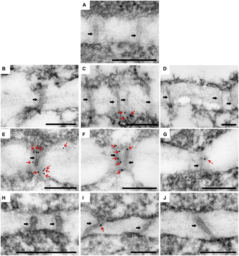
FIGURE 6.
Immunolocalization of β-1, 3-glucan (callose) in the PDs in SAM of Argyranthemum. (A) Negative control. (B–D) Plasmodesmata (PD) of CSVd infected ‘Yellow Empire’, in zone 1, 2, 3, respectively. (E–G) PD of CSVd infected ‘Border Dark Red’, in zone 1, 2, 3, respectively. (H–J) PD of healthy ‘Border Dark Red’, in zone 1, 2, 3, respectively. Immunogold particles show β-1, 3-glucan (callose) accumulation in PD. Black arrows indicate PD. Red arrows indicate immunogold particles (callose). Scale bars of (A–J) are 200 nm.

