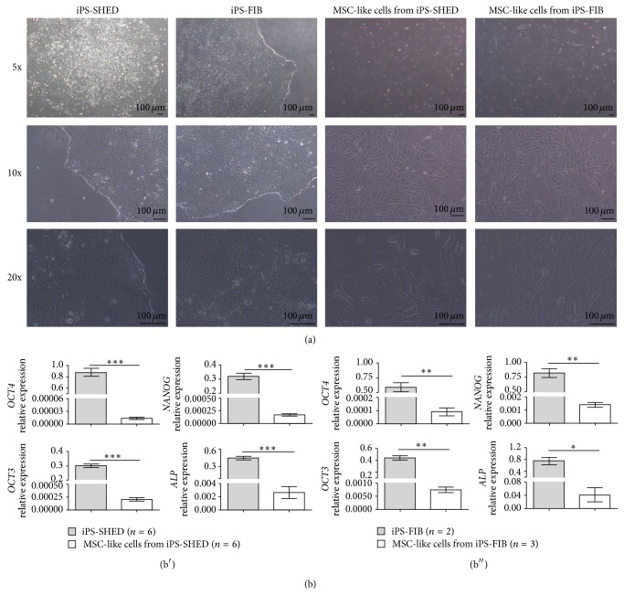
Figure 1.
(a) Morphology of undifferentiated hiPSC colonies cultured on matrigel and MSC-like cells from iPS-SHED and iPS-FIB after 12 days of in vitro mesenchymal induction. Scale bar = 100 um. (b) Real-time quantitative PCR analysis of pluripotency markers in undifferentiated hiPSCs ((b′) SHED and (b′′) fibroblasts) and in MSC-like cells from iPS-SHED and from iPS-FIB. ACTB, TBP, and HMBS were used as endogenous controls. Values represent means +/− SD, P < 0.05 (*), P < 0.01 (**), and P < 0.001 (***).

