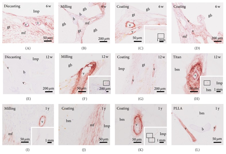
Figure 7.
Location of blood vessels by anti-smooth muscle actin (ASMA) immunohistochemistry. Vessels (v) and myofibroblast (mf) are identified by their red staining in the granulation tissue (gt) located between the gas voids (gb) and at the implant (Imp) interface at the 6-week (w) follow-up time (A–D). After 12 weeks the amount of ASMA staining was reduced (E–H). In the PLLA (L) and titanium (H) controls no myofibroblast was labeled and vessels were mainly found in erosions lacunae and bone marrow (bm). The amount of myofibroblasts was increasingly reduced in the samples with Mg implants. Only a limited number of blood vessels were found mainly in bone marrow. Insets in (C), (F), (H), (I), and (K) overviews.

