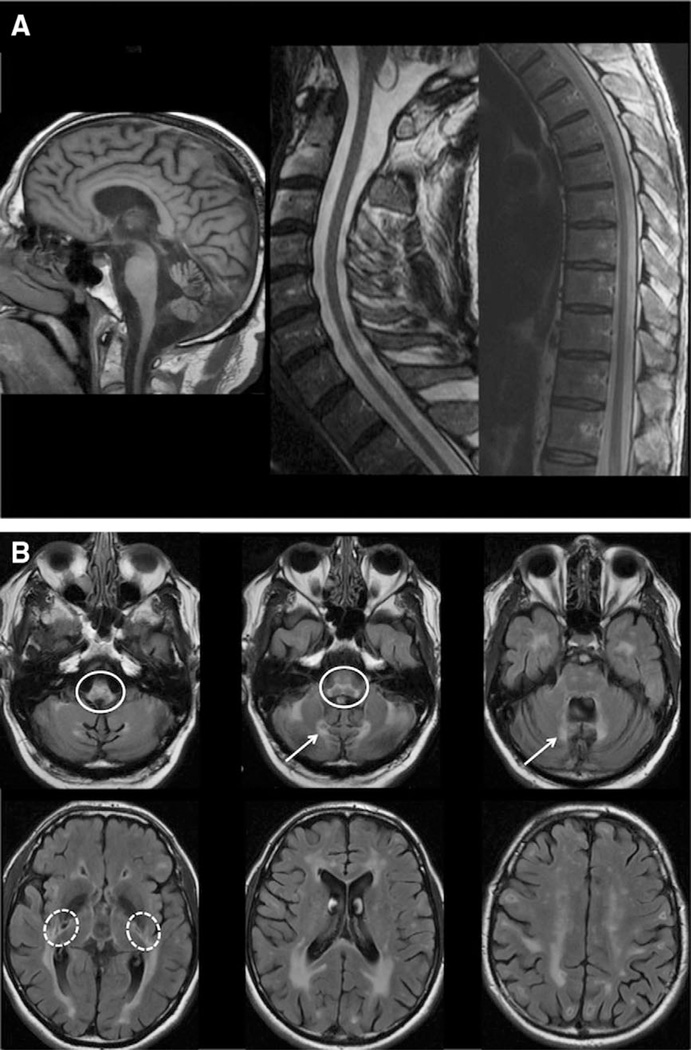
Figure 2.
Typical cerebral and spinal pattern is shown in adult polyglucosan body disease patients. (A) T1 sagittal scans show important medullary and spinal atrophy and mild vermian atrophy. (B) Fluid attenuated inversion recovery axial scans show hyperintense white matter abnormalities in the periventricular regions, with occipital predominance, the external capsule and the posterior limb of the internal capsule (dashed circles), the medial edges of the inferior and middle cerebellar peduncles (arrows), and the pyramidal tracts and medial lemniscus of the medulla and pons (plain circles).

