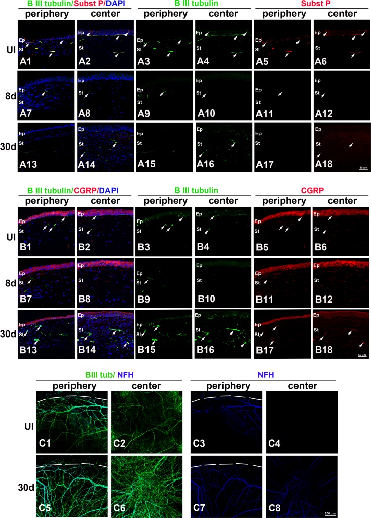
Figure 2.
HSV-1 infection results in loss of corneal sensory fibers and a deficient or abnormal reinnervation process. Mouse eyes were infected with 103 PFU HSV-1 or left UI as controls. At 8 and 30 days pi, corneas were processed for IHC costaining with β III tubulin (green) with SP (red), CGRP (red), or NFH (blue) antibodies. (A1–A18) Sagittal frozen sections of corneas costained with β III tubulin and SP. In the UI cornea, both periphery (A1, A3, A5) and center (A2, A4, A6) showed β III tubulin+ nerves in the stroma and rich subbasal innervation penetrating the layers of epithelial cells that colocalized with expression of SP (white arrows). Loss of sub-basal innervation was observed at 8 days pi (A7–A12) with remaining peripheral nerves that did not colocalize with SP (A7, A9, A11). The reinnervation of the cornea at 30 days pi (A13–A18) involved poor sub-basal reinnervation and hyperinnervation of the central stroma without recovery of SP+ signal (white arrows). (B1–B18) Sagittal frozen sections of corneas costained with β III tubulin and CGRP. In the UI cornea, CGRP expression was found mostly within the epithelial cells (B1–B6) and was occasionally associated with nerves at the sub-basal and stromal locations. At 8 days pi, CGRP continued to be expressed in the epithelium and was not associated with remaining nerves at the peripheral cornea (B7–B12). At day 30 pi, CGRP reactivity at the central epithelium region was largely lost, whereas some of the nerves that reinnervated the stroma colocalized with CGRP expression (B13–B18). (C1–C8) Corneal whole mounts show hyper-reinnervation at the center of the cornea at 30 days pi that included the abnormal presence of NFH+ nerves. DAPI, nuclei counterstain in blue; Ep, epithelium; St, stroma. Figure represents n = 3 mice/time point or UI controls from two independent experiments.

