Figure 1.

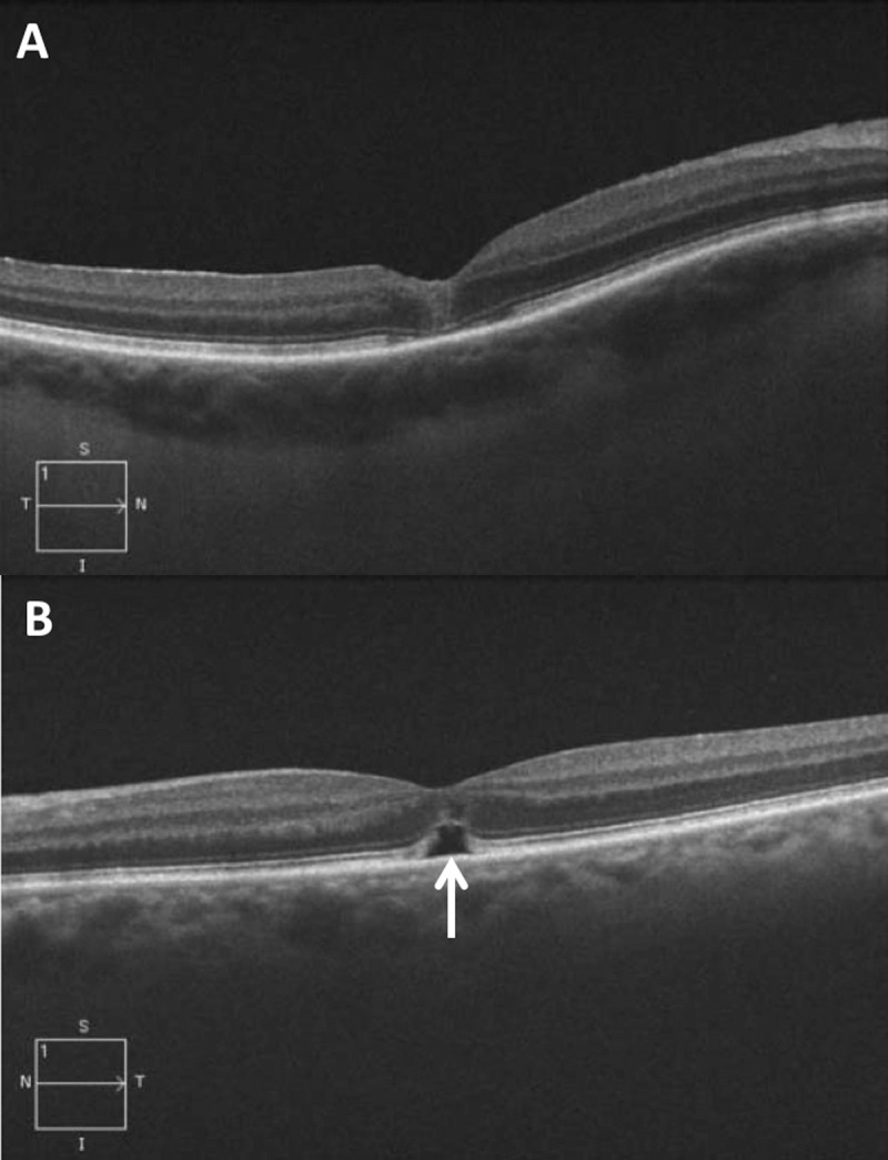
Optical coherence tomography B-scans 2 weeks after successful MH repair. (A) Case example without subfoveal fluid after surgery. (B) Case example with persistent subfoveal fluid (white arrow).

Optical coherence tomography B-scans 2 weeks after successful MH repair. (A) Case example without subfoveal fluid after surgery. (B) Case example with persistent subfoveal fluid (white arrow).