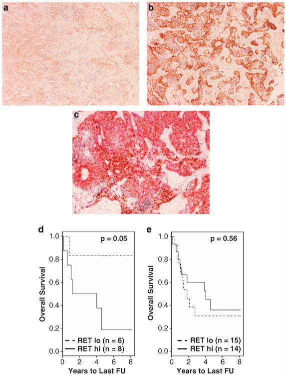
Figure 5.

RET protein expression by IHC. RET staining in fatal adenocarcinoma was typically much less intense in ASCL1− (a) than in ASCL1+ (b) tumors. (c) Co-IHC of ASCL1 (nuclear brown staining) and RET (cytoplasmic red staining) identified areas with overlapping expression of the two proteins. (d) KM plot of 14 ASCL1+ AD samples indicates a significant association with OS (P = 0.05). (e) When samples were not stratified by the ASCL1 expression, RET IHC was not significant in predicting OS.
