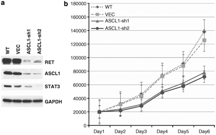
Figure 6.

In vitro analysis of ASCL1 and RET in HCC1833 AD cells. (a) Wild-type cells (WT) express high levels of ASCL1 and RET. Knocking down of ASCL1 by stable shRNA transfection (ASCL1-sh1 and ACSL1-sh2) resulted in significant reduction of RET protein levels in these cells compared with cells transfected with empty vector (VEC), suggesting that ASCL1 acts upstream of RET. Also, STAT3 levels were reduced in ASCL1-sh cells, suggesting potential activation of the JAK/STAT3 pathway. (b) ASCL1-sh cells had markedly reduced proliferation rates compared with VEC and WT cells.
