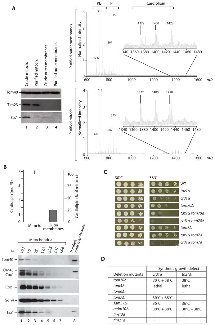
Figure 1. Outer membrane lipids and genetic interaction of cardiolipin mutants with tom/sam mutants.
(A) Purified yeast mitochondrial outer membrane vesicles were analyzed by immunoblotting (left panel) and liquid chromatography/mass spectrometry (right panels). PE, phosphatidylethanolamine; PI, phosphatidylinositol; Sss1, endoplasmic reticulum protein.
(B) Cardiolipin was quantified [11] using tetramyristoyl cardiolipin as internal standard. Data from two independent outer membrane preparations are represented as mean +/− range. The purity of outer membrane vesicles was determined by a Western blot titration of outer membrane proteins (Tom40, OM45) and inner membrane proteins (Cox1, Sdh4).
(C and D) Genetic interactions, synthetic growth defects. Cells were grown at 30°C in liquid YPD to the early stationary phase, serially diluted, spotted on YPD plates and incubated at the indicated temperatures. Synthetic growth defects of double deletion mutants are indicated; ‘-‘ no synthetic growth defect.

