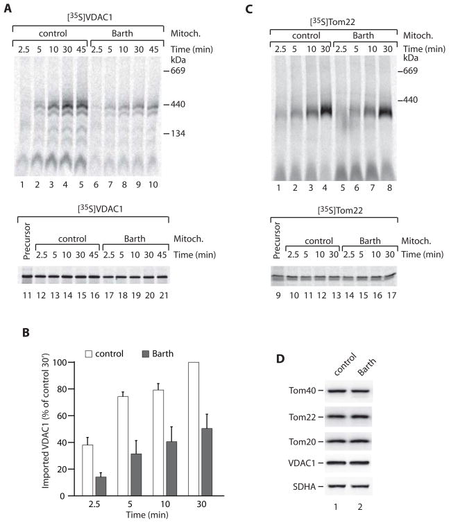
Figure 4. Assembly of outer membrane proteins in Barth syndrome mitochondria.
(A–C) Mitochondria isolated from control or Barth syndrome patient lymphoblasts were incubated at 37°C (min) in the presence of [35S]VDAC1 or [35S]Tom22. Following import, samples were analyzed by blue native electrophoresis (upper panel) and SDS-PAGE (lower panel) and autoradiography. A sample of reticulocyte lysate (representing 50% of added precursor/import) is also shown on the SDS-PAGE gel. For B, assembled VDAC1 was quantified from four independent experiments (three for 2.5 min). Data are represented as mean +/− SEM.
(D) Control and Barth syndrome mitochondria were analyzed by SDS-PAGE and immunoblotting. SDHA, 70 kDa subunit of succinate dehydrogenase.

