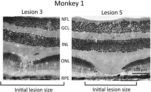
Fig. 4.
Histological appearance of two lesions in monkey 1. Images show the range of observed gap in photoreceptor inner and outer segments; no gap in lesion 3 versus a 120 um gap in lesion 5. Of the 6 lesions examined in monkey 1, only lesion 5 showed a substantial gap in photoreceptor inner and outer segments, all other lesions showed little or no gap. The outer nuclear layer (ONL), which contains the soma of all photoreceptor cells, was interrupted by all lesions, although the gap in the ONL was smaller in all cases (see Table 1) than the initial lesion size as measured by fundus photography (Table 1). Ganglion cell layer (GCL) and inner nuclear layer (INL) showed no morphological changes at the location of lesions, nor was the nerve fiber layer (NFL) affected. We found no alteration of Muller cell morphology in neighboring vimentin stained sections. The RPE cell monolayer appeared intact across all lesions, although melanin pigment granules are reduced in some lesions (seen here in lesion 5, but not in lesion 3). Neighboring sections showed intact lipofuscin content in the monolayer of RPE cells across all lesions. Hematoxylin and eosin stain, 20×.

