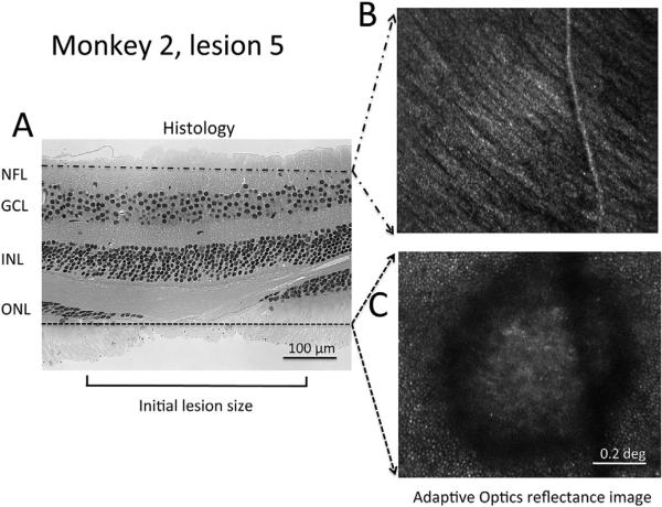
Fig. 5.
Histology and AO images of lesion 5 in monkey 2. Comparison of a histological section through lesion 5 in monkey 2 to two images from a through-focus stack of invivo AO images of the same region obtained approximately 2 months before euthanasia. A. Section through lesion 5 that illustrates no apparent change in inner retinal layers (upper part of the image) but interruption of the outer nuclear layer (ONL), and the presence of photoreceptor inner and outer segments extending farther into the lesion that the ONL. B. In vivo adaptive optics image showing superficial nerve fiber layer, and C. photoreceptors surrounding the lesion. Hematoxylin and eosin stain, 20×.

