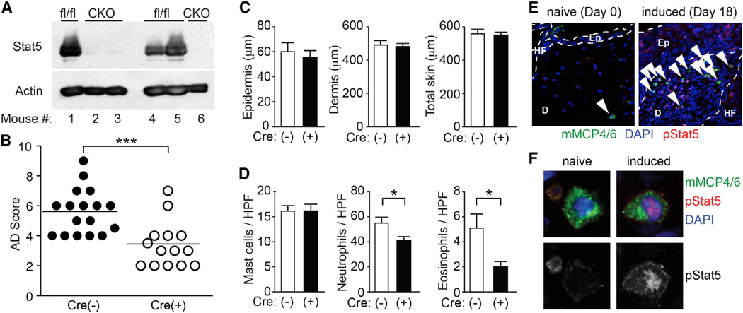
Figure 4. Stat5 in Mast Cells Regulates Der f/SEB-Induced Dermatitis.
Dermatitis was induced with Der f/SEB in MCΔStat5 mice [CKO or Cre(+)] and their floxed control [fl/fl or Cre(−)] mice. *p < 0.05, ***p < 0.001 by Student’s t test.
(A) Western blot analysis of Stat5 in mast cells derived from neonatal skin of MCΔStat5 (CKO) and control (fl/fl) mice.
(B) AD scores accumulated from four separate experiments using three to five mice per group.
(C) Thicknesses of epidermis and dermis after Der f/SEB (D/B) treatment.
(D) Histologic analysis of Der f/SEB-induced dermatitis. Data represent mean ± SEM.
(E and F) Skin sections of naive and Der f/SEB-induced (6 hr after fourth induction) dermatitis in WT mice were stained for phospho-Stat5, mMCP4, and mMCP6. Arrowheads indicate pStat5-positive mast cells. Ep, epidermis; D, dermis; HF, hair follicle. Representative images of mast cells from three experiments are shown in (E) and (F). See also Figure S4.

