
Figure 2.
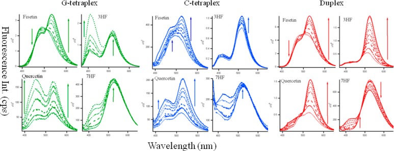
Fluorescence emission spectra of fisetin (λex = 370 nm), quercetin (λex = 370 nm) 3-hydroxyflavone (3HF, λex = 350 nm) and 7-hydroxyflavone (7HF, λex = 350 nm) (all ∼10 μM) in the presence of increasing concentration of G4 (green), C4 tetraplex (blue), and duplex DNA (red) at ∼0–25 μM), (0····, 2 − - - −, 5 − − −, 10 −··−··−, 15 −·−·−, 20 - - -, 25 — μM). Slit widths were 3,3 for 3HF and fisetin and 3,5 for quercetin and 7HF.