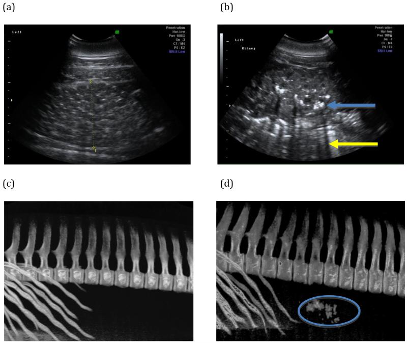
Figure 1.
Representative renal images from study animals. (a) Dorsal plane renal sonogram of the left kidney from an animal with no stones. (b) Dorsal plane renal sonogram of the left kidney of an animal with a heavy stone burden. Hyperechoic foci (blue arrow) with acoustic shadowing (yellow arrow) are characteristic of calculi on ultrasound examination. (c) Representative three-dimensional reconstruction of CT data that include the caudal ribs and lumbar vertebral bodies, evaluated specifically for mineral density in the region of the kidneys. No mineral densities were detected in this non-stone former. (d) Numerous mineral densities in the kidneys were detected in the reconstructed CT data of a stone-forming dolphin (oval).

