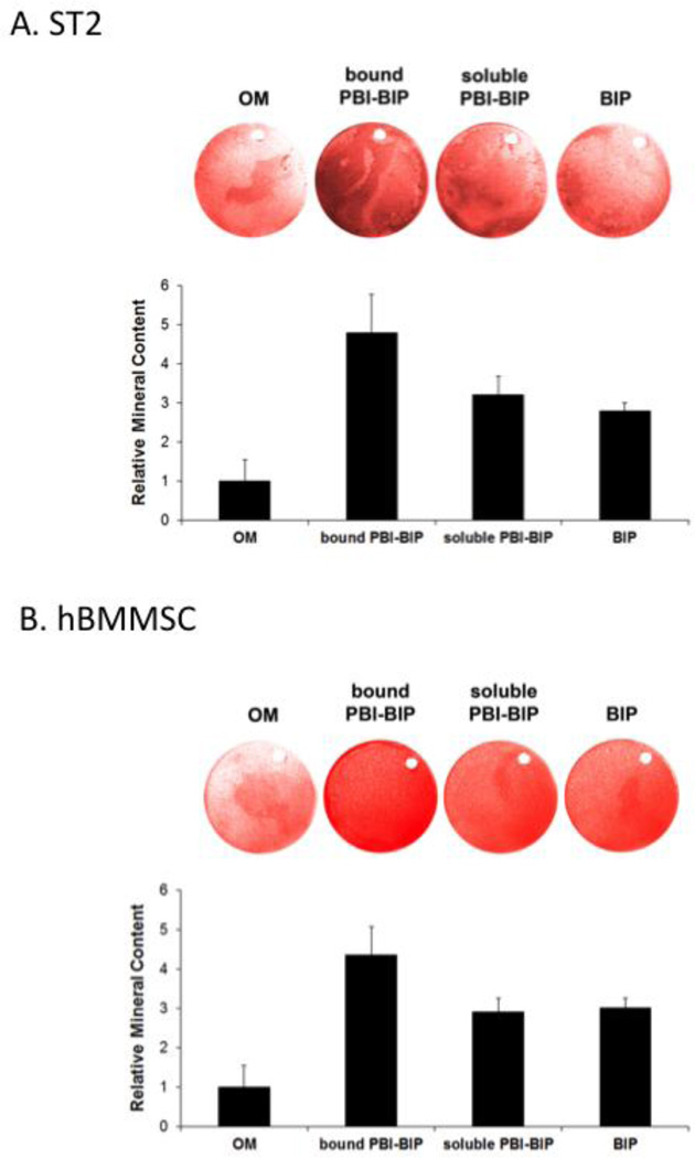
Figure 3. Differentiation of cells grown on PBI-BIP chimeric peptide-bound implant surfaces.
Mouse ST2 cells (A) or hBMMSCs (B) were grown on titanium discs for two weeks (ST2) or four weeks (hBMMSCs). Mineral deposition was assayed with Alizarin Red S staining and quantified. “OM,” osteogenic medium alone; “bound PBI-BIP,” PBI-BIP preloaded onto Ti disc in OM; “soluble PBI-BIP,” PBI-BIP in OM; “BIP,” BIP in OM.

