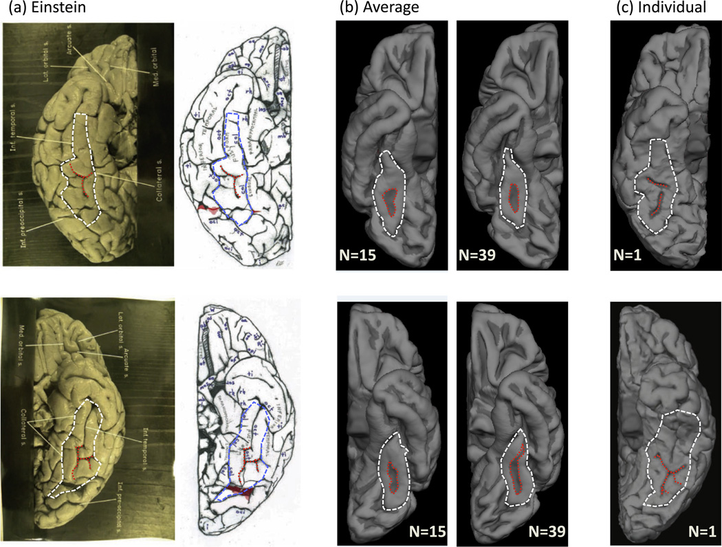
Fig. 1.
Einstein’s sulcal patterning on the fusiform gyrus (FG) is abnormal relative to a group average, but reflective of a subset of individuals. (a) Original (left) and schematic (right) images from Falk et al. (2013) for the right (top) and left (bottom) hemispheres of Einstein. Outlines of the FG (white/blue) and the mid-fusiform sulcus (MFS; red) have been added. (b) Average inflated cortical surfaces from two different independent sets of ‘typical’ brains. Left: N = 15; Right: N = 39 (FreeSurfer template). Red: MFS. White: FG. (c) Example pial surfaces from individual subjects for the right (top) and left (bottom) hemispheres. Red: MFS. White: FG. Einstein’s MFS in both hemispheres deviates from the group, but resembles sulcal patterns reflective of a minority of individuals. (For interpretation of the references to color in this figure legend, the reader is referred to the web version of this article.)

