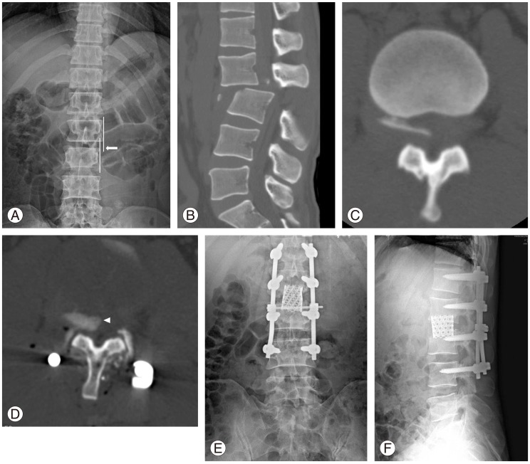
Fig. 3.
A 47-year-old male patient with fracture-dislocation injury. (A) Lateral translation of L3 compared to L2 on anteroposterior radiographs (arrow) right after the injury. (B) Posterior translation of L3 to L2 on sagittal computed tomography (CT) scan right after the injury. (C) Bony fragment within the spinal canal on preoperative axial CT scan. (D) Bony fragments were not reduced after the reduction and posterior instrumented fusion (arrow head). (E, F) Anteroposterior and lateral radiographs after removal of bony fragments and fusion with cages through the anterior approach.

