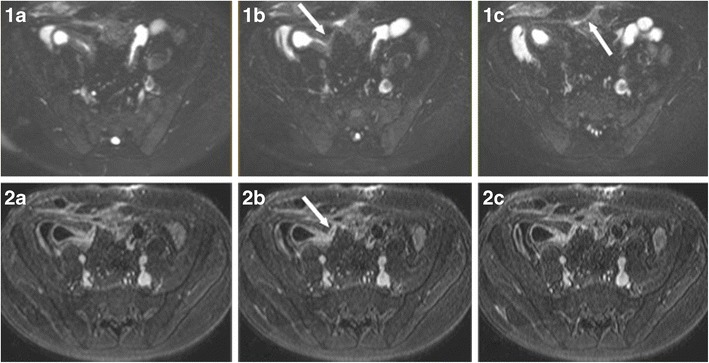
Fig. 5.

Ileocutaneous fistula in a 13-year-old male. T2 fat-saturated (1a–c) and T1 contrast-enhanced (2a–c) images. Direct visualisation of the fistula (arrows) is feasible as it shows avid contrast enhancement and contents of increased T2 signal, suggestive of enteric material
