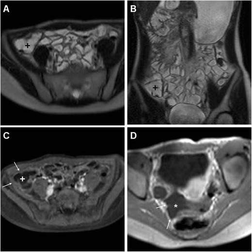
Fig. 3.

A 16-year-old female with intermittent low-grade fever, failure to thrive and weight loss 15 weeks after LA for uncomplicated AA. Considering the patient’s young age, to avoid use of ionising radiation investigation was carried out by means of MR enterography including peroral bowel distension with diluted polyethylenglycol solution. Axial (a) and coronal (b) T2-weighted images show optimal ileal and caecal (+) distension by intraluminal fluid, without residual inflammatory fat stranding and appreciable abscess collection. After intravenous gadolinium-based contrast, T1-weighted images with (c) and without (d) fat suppression exclude abnormal enhancement of the peritoneal serosa (thin arrows), with minimal fluid (* in d) in the peritoneal cul-de-sac. The patient did not require further treatment
