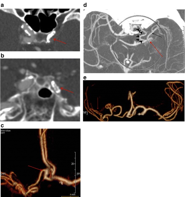
Fig. 11.

a Axial CT image at the level of the cavernous carotids shows occlusion of the left ICA (red arrow) due to atherosclerotic disease. b Coronal reconstruction at the level of the cavernous carotids verifies the occlusion of the left ICA (red arrow) as well as atherosclerotic disease. c CTA (VRT 3D reconstructions) shows the accessory MCA as a vessel coming from the A2 segment of the ipsilateral anterior cerebral artery (red arrow). d Axial MIP image shows the course of the AccMCA (black arrowheads), the anastomotic network (moyamoya type) at the level of the left mid M1 segment (red arrow) as well as the patent peripheral left MCA (black arrow). e CTA (VRT 3D reconstructions) reveals the same configuration and patent peripheral MCA (red arrow)
