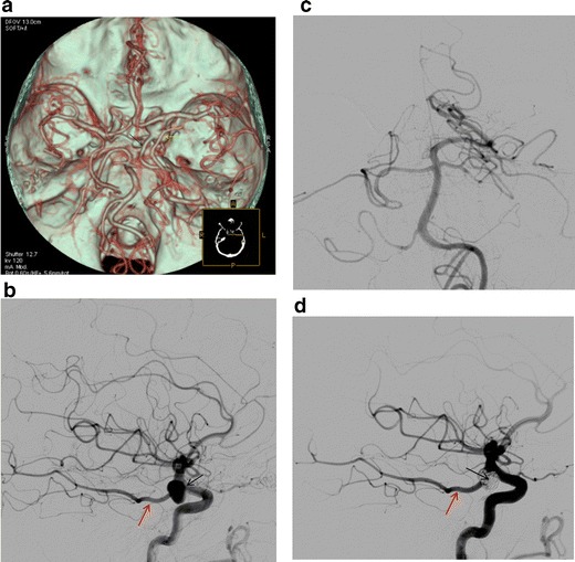
Fig. 12.

a CTA (VRT reconstructions) shows a right-sided foetal Pcom (black arrow) with a large aneurysm at the origin of the vessel (yellow arrow). b Digital subtraction angiography (lateral view) of the right ICA shows a foetal Pcom (red arrow) and large aneurysm at the origin of the vessel (black arrow), which cannot be compromised. c Digital subtraction angiography (AP view) of the left vertebral artery. The posterior cerebral artery on the right is not opacified (definition of a foetal Pcom). b Post-embolisation digital subtraction angiography (lateral view) of the right ICA shows good patency of the foetal Pcom (red arrow) and complete obliteration of the aneurysmal sac (black arrow)
