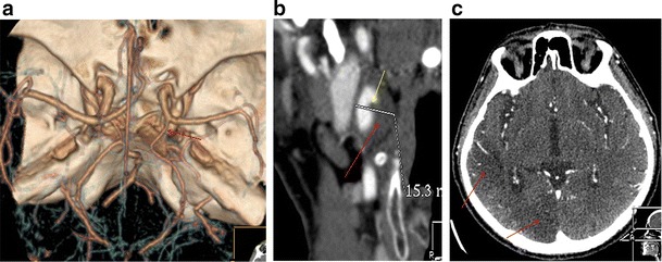
Fig. 13.

a CTA (VRT reconstructions) shows a right-sided foetal Pcom (red arrow). b CT oblique MPR image shows an increased wall thickness (red arrow) and intimal flap (yellow arrow) of the dissected right ICA. c Axial CT image at the level of the third ventricle shows right-sided occipital and posterior parietal lobe infarcts (red arrows)
