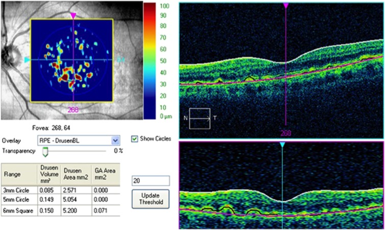
Figure 1.
The Cirrus automated algorithm provides quantitative measurements of macular drusen area and volume within a 3 mm CC and a 3–5 mm surrounding PR. The RA measurements are provided for the entire 6 × 6 mm2 area. It uses the RPE geometry (black line) and compares this segmentation map with a virtual map of the RPE free of deformations (pink line). Using these two maps, the algorithm creates an elevation map that permits measurement of drusen area and volume.

