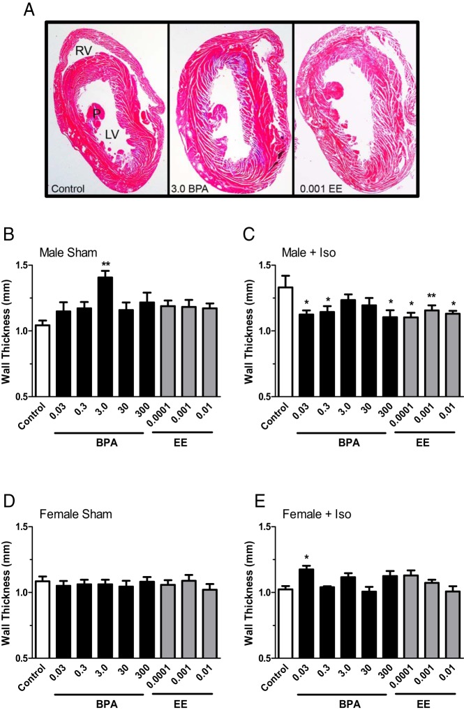
Figure 3.
The impact of BPA and EE on LV wall thickness and effects of exposures on the response to isoproterenol in male and female CD-1 mice exposed to BPA or EE. A, Representative photomicrographs of hematoxylin and eosin-stained sections of male hearts from the control, 3.0 ppm BPA and 0.001 EE exposure groups. LV, right ventricle (RV), and papillary muscle (P) are indicated. In sham BPA-exposed males (B), a significant increase in LV wall thickness was observed for the 3.0 ppm exposure group. Compared with sham control males, Iso increased LV wall thickness, an effect that was reversed in each BPA and EE exposure group except for the 3.0 and 30 ppm BPA exposure groups (C). In females exposure to BPA or EE did not affect LV wall thickness in sham-treated females (D). In females from the lowest BPA group (0.03), a significant increase in LV wall thickness was observed in response to Iso (E). Data are presented as group mean of wall thickness in millimeters ± SEM. The level of statistical significance determined from a two-way ANOVA, and Bonferroni's multiple comparison tests are indicated with asterisks.

