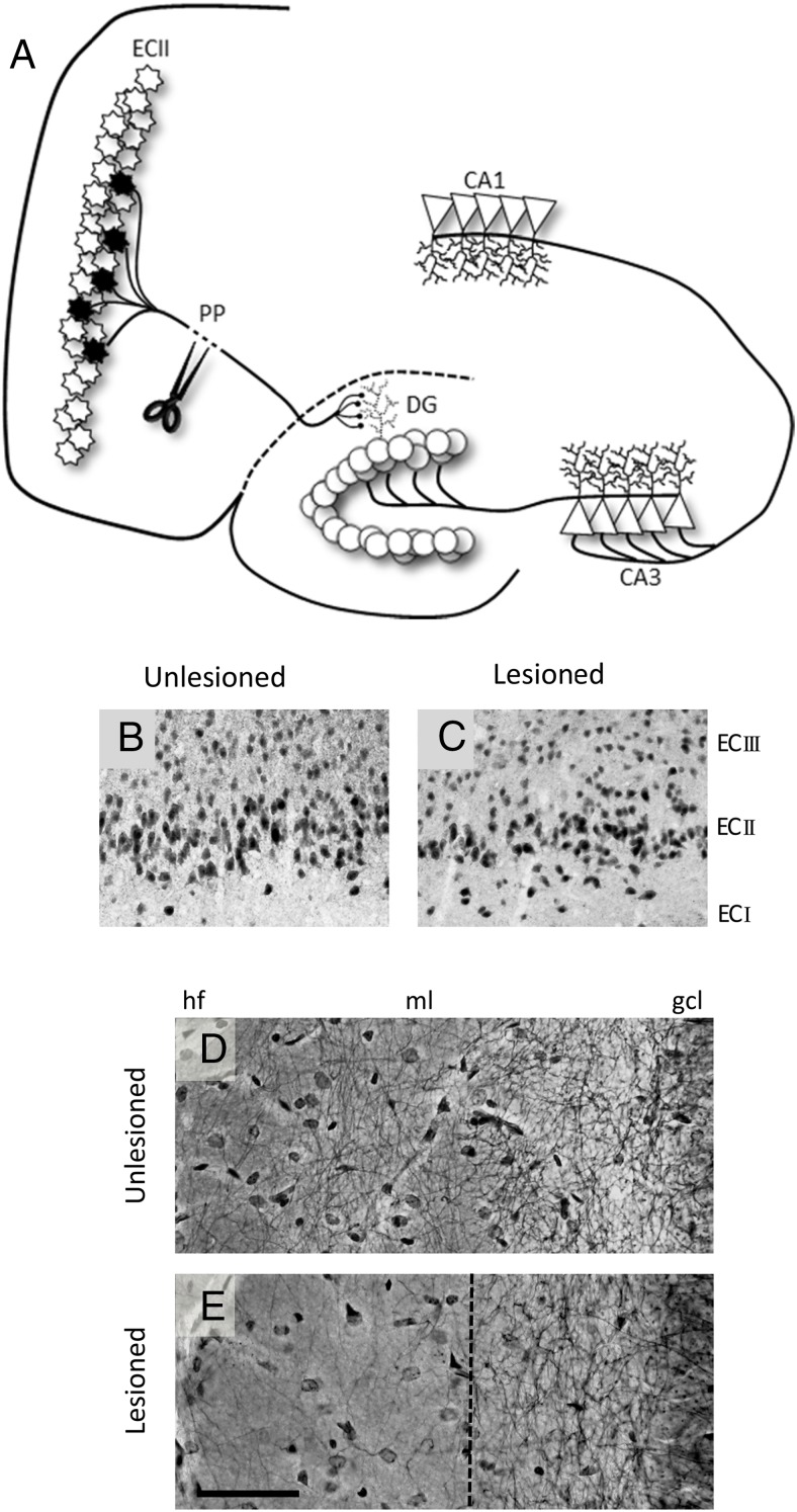
Figure 1.
Unilateral entorhinal cortex lesion model. A, Schematic representation of the entorhinal cortex/perforant path lesion model. Perforant path (PP) axons arise from the distinctly laminated layer II entorhinal cortex neurons (ECII) and terminate on the dendrites of the granule cells in the molecular layer of the DG. Unilateral lesion of the perforant path results in ipsilateral degeneration of axotomized ECII neurons, evidenced by reduced NeuN immunoreactive cells in the ECII region of the lesioned (ipsilateral, B) vs unlesioned hemisphere (contralateral, C). Neurite fiber density in the DG molecular layer, as assessed by Holmes staining, is reduced after the lesion (unlesioned, D; lesioned, E) but shows compensatory regrowth. Compensatory sprouting arises from reinnervation from multiple pathways including commissural/associational afferents, septohippocampal afferents, contralateral EC afferents, and local interneurons (71). Scale bar, 50 μm. hf, hippocampal fissure; ml, molecular layer; gcl, granule cell layer.

